Estudio de la estructura macroscópica y microscópica glándula del seno infraorbitario en la oveja Tuj
Resumen
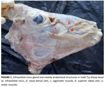
El seno infraorbitario (glándula infraorbitaria-glándula preorbitaria) está ubicado en el aspecto lateral del cráneo, rostroventral al ojo, dentro de la fosa infraorbitaria. Algunas glándulas cutáneas están diferenciadas y especializadas. En el presente estudio, se buscó examinar la estructura macroscópica y microscópica del glándula seno infraorbitario, una de estas glándulas. La distancia entre el foramen infraorbitario y el seno infraorbitario fue de 47.35±0.57 mm en el lado derecho y de 53.80±1.27 mm en el izquierdo. La longitud del lado izquierdo del seno infraorbitario en la oveja Tuj fue de 25.32±2.21 mm, la anchura de 16.21±2.83 mm y la profundidad de 1.23±1.80 mm. El seno infraorbitario de la oveja Tuj estaba compuesto por la epidermis y la dermis. La epidermis estaba compuesta por epitelio escamoso estratificado queratinizado. Se observaron numerosas glándulas sebáceas, algunas glándulas apocrinas, fibras musculares y folículos pilosos en la dermis. Las glándulas sebáceas se ubicaban en la región superficial y las apocrinas en la profundidad de la dermis. Se observaron numerosas gotitas de lípidos dentro de las células de las glándulas sebáceas. Las células mioepiteliales y la membrana basal de las glándulas apocrinas y las fibras musculares dieron positivo a PAS. En conclusión, en el estudio presentado, se determinó la estructura macroscópica y microscópica del seno infraorbitario en ovejas Tuj. Esta región también es clínicamente importante porque está cerca del nervio infraorbitario que pasa por el agujero infraorbitario (área de anestesia) y el seno infraorbitario está ubicado topográficamente en esta región. Se cree que este estudio respaldará los estudios científicos relacionados con la piel y sus apéndices.
Descargas
Citas
Demiraslan Y, Orhun-Dayan M. Veteriner Sistematik Anatomi. 1st. ed. Ankara, Turkey: Atlas Kitabevi; 2021.
Demiraslan Y, Orhun-Dayan M. VETKA-Veteriner Klinik Anatomi. 1st. ed.Ankara, Turkey: Nobel Akademik Yayincilik; 2023.
König HE, Liebich HG. Veteriner Anatomi (Evcil Memeli Hayvanlar). 3rd. ed.Turkey: Medipres; 2022.
Haziroglu RM, Cakir A, editors. Veteriner Anatomi Konu Anlatımı ve Atlas. 4th. ed. Ankara: Güneş Tip Kitabevleri; 2017.
Moawad UK. Morphological, histochemical and morphometric studies of the preorbital gland of adult male and female egyptian native breeds of sheep (Ovis aries).Asian J. Anim. Vet. Adv. [Internet]. 2016; 11(12):771-782. doi: https://doi.org/p73r. DOI: https://doi.org/10.3923/ajava.2016.771.782
King N, Kumar AHS, Kilroy D. Ovine infraorbital (preorbital) pouch: an unusual presentation in a male lamb.Biol. Eng. Med. Sci. Rep. [Internet]. 2018; 4(1):01-02. doi: https://doi.org/p73s. DOI: https://doi.org/10.5530/bems.4.1.1
Rajagopal T, Archunan G. Histomorphology of preorbital gland in territorial and non-territorial male blackbuck Antelope cervicapra, a critically endangered species.Biol. [Internet]. 2011; 66:370-378. doi: https://doi.org/cj22wj. DOI: https://doi.org/10.2478/s11756-011-0015-4
Dursun N. Veteriner Anatomi II. 12th. ed. Ankara: MedisanPublishing House; 2008.
Delmann HD, Brown EM. Textbook of Veterinary Histology. 2nd. Ed. Philapelphia: Lea & Febiger; 1981.
Atoji Y, Yamamoto Y, Suzuki Y. Infraorbital glands of a male formosan serow (Capricornis crispus swinhoei). Eur. J. Morphol. [Internet]. 1996; 34(2):87-94. doi: https://doi.org/fpczwb. DOI: https://doi.org/10.1076/ejom.34.2.87.13015
Ceaceroa F, Pluhácek J, Komárková M, Zábranský M. Pre- orbital gland opening during aggressive interactions in Rusa deer (Rusa timorensis). Behav. Proces. [Internet]. 2015; 111:51–54. doi: https://doi.org/f62ppw. DOI: https://doi.org/10.1016/j.beproc.2014.11.017
N.A.V. International Committee on Veterinary Gross Anatomical Nomenclature. Nomina Anatomica Veterinaria (I.C.V.G.A.N.). 6th. ed. World Association of Veterinary Anatomists: Hanover (Germany), Ghent (Belgium), Columbia, MO (U.S.A.), Rio de Janeiro (Brazil); 2017.
Liu M, Yang Y, Li M, Cai Y, Zhao S, Wang X. Comparison study of morphology andvascular casts between plateau- type Tibetan sheep andlow-altitude small-tailed Han sheep testes. Anat. Histol. Embryol. [Internet]. 2022; 51(4):524–532. doi: https://doi.org/p74r. DOI: https://doi.org/10.1111/ahe.12822
Adnyane IKM, Zuki ABZ, Noordin MM, Wahyuni S, Agungpriyono S. Morphological study of the infraorbital gland of the male barking deer, muntiacus muntjak. Afr. J. Biotechnol. [Internet]. 2011; 10(77):17891-17897. Available in: https://goo.su/TOR4k. DOI: https://doi.org/10.5897/AJB10.2634
Clutton-Brock T, Guinness FE, Albon SD. Red Deer, Behaviour and Ecology of Two Sexes. 1st. ed. Chicago, IL: University of Chicago Press; 1982.
Bartos L. Some observations on the relationship between preorbital gland opening and social interactions in red deer. Aggress. Behav. [Internet]. 1983; 9(1):59–67. doi: https://doi.org/dvpzk2. DOI: https://doi.org/10.1002/1098-2337(1983)9:1<59::AID-AB2480090108>3.0.CO;2-F
Hatlapa HH. Zur biologischen Bedeutung des praorbital Organs beim Rotwild, Pragung, Individualgeruch, Orientierung. Berl. Munch. Tierartzl. Wochenschr. 1977; 90:100–104.
Wolfel H. Zur Jugendentwicklung, Mutter-Kind-Bindung und Feindvermeidung beim Rothirsch (Cervus elaphus) II. Z. Jagdwissensch. [Internet]. 1983; 29:197–213. doi: https://doi.org/dgdq8t. DOI: https://doi.org/10.1007/BF02243645
Bartos L, Víchová J, Lancingerová J. Preorbital gland opening in red deer (Cervus elaphus) calves: Signals of hunger?. J. Anim. Sci. [Internet]. 2005; 83(1):124–129. doi: https://doi.org/p746. DOI: https://doi.org/10.2527/2005.831124x
Eurell JA, Frappier BL. Dellmann’s Textbook of Veterinary Histology. 6th. ed. Iowa (USA), Oxford (UK): Blackwell Publishing; 2006.
Gernek WH, Cohen M. The micromorphology of the glands of the infra-orbital cutaneous sinus of the Steenbok (Raphıcerus Campestrıs). Onderstepoort J. Vet. Res. [Internet]. 1978; 45(2):59-66.
Mohamed R, Driscoll M, Mootoo N. Clinical anatomy of the skull of the Barbados Black Belly sheep in Trinidad. Int. J. Curr. Res. Med. Sci. [Internet]. 2016; 2(8):8-19. Available in: https://goo.su/fQ9X.