
Análisis histológico del epidídimo y los testículos en la dinámica de la ontogénesis postnatal desde un mes hasta la pubertad de los corderos Ouled Djellal
Resumen
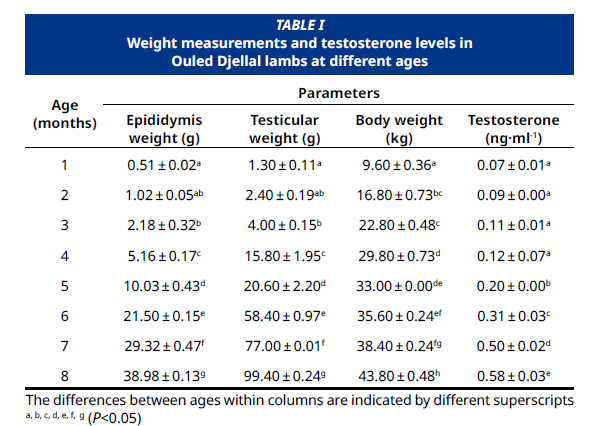
El objetivo del estudio fue analizar la ontogenia del epitelio epididimario y seminífero en el testículo de los corderos Ouled Djellal a lo largo del desarrollo postnatal hasta la pubertad. Se utilizaron un total de 24 corderos Ouled Djellal, con edades comprendidas entre 1 y 8 meses, seleccionando tres corderos en cada mes de edad. Los corderos fueron castrados quirúrgicamente mensualmente, y se realizó histología de rutina junto con histomorfometría en los tejidos epididimarios y testiculares procesados utilizando el software AxioVision Rel 4.6. El análisis estadístico reveló que el peso testicular aumentó a un ritmo acelerado entre los 4 y los 8 meses postnatales, coincidiendo con modificaciones significativas en la histología testicular. Estas modificaciones abarcaron incrementos significativos en la medida de los túbulos seminíferos y un incremento en los niveles de testosterona. Se encontró que el diámetro ductal, el diámetro luminal y la altura del epitelio eran estadísticamente diferentes (P<0.05) entre las regiones caput, corpus y cauda del epidídimo, con los niveles máximos registrados durante la pubertad. A los 1 y 2 meses de edad, había dos tipos de células presentes en el epitelio seminífero: células de soporte y gonocitos. A los 3 meses de edad, se formaron los primeros espermatogonios a partir de gonocitos, coincidiendo con el peso promedio de los testículos alcanzando 4 g. La espermatogénesis se inició cuando los gonocitos sufrieron mitosis, dando lugar a progenitores que se diferenciaron en espermatogonios. A los 4 y 5 meses de edad, se observó inicialmente un lumen y espermatocitos primarios (espermatocitos I) en los túbulos seminíferos. A los 6 meses de edad, espermatocitos secundarios y espermatides redondas formaron una única fila dentro de los túbulos seminíferos. A los 7 y 8 meses de edad, todas las generaciones de células madre germinales se encontraban en los seminiferous tubules. A los 8 meses de edad, se observaron espermatozoides en varios segmentos del epidídimo en la especie Ouled Djellal, dando inicio a la etapa de pubertad.
Descargas
Citas
Jumaa AJ, Kassim WY. The influence of age, gender and weight on sex hormone levels and histological development of reproductive organs in Arabi sheep. Al–Mustansiriyah J. Sci. [Internet]. 2023; 33(5):27–32. doi: https://doi.org/n4wp DOI: https://doi.org/10.23851/mjs.v33i5.1309
Herrera–Alarcón J, Villagómez–Amezcua E, González–Padilla E, Jiménez–Severiano H. Stereological study of postnatal testicular development in Blackbelly sheep. Theriogenology [Internet]. 2007; 68(4):582–591. doi: https://doi.org/dj38vd DOI: https://doi.org/10.1016/j.theriogenology.2007.01.020
Sarlós P, Egerszegi I, Balogh O, Molnár A, Cseh S, Rátky J. Seasonal changes of scrotal circumference, blood plasma testosterone concentration and semen characteristics in Racka rams. Small Rumin. Res. [Internet]. 2013; 111(1–3):90–95. doi: https://doi.org/f4sf98 DOI: https://doi.org/10.1016/j.smallrumres.2012.11.036
Bielli A, Genovese P, Ungerfeld R, Katz H. Histology of lamb epididymal development. Anat. Histol. Embryol. [Internet]. 2007; 36(6):437–441. doi: https://doi.org/bbj2k8 DOI: https://doi.org/10.1111/j.1439-0264.2007.00782.x
Ibrahim AA, Aliyu J, Ashiru RM, Jamilu M. Biometric study of the reproductive organs of three breeds of sheep in Nigeria. Int. J. Morphol. [Internet]. 2012 [cited 24 Jul. 2024]; 30(4):1597–1603. Available in: https://n9.cl/v0nj5 DOI: https://doi.org/10.4067/S0717-95022012000400053
Kangawa A, Otake M, Enya S, Yoshida T, Shibata M. Histological development of male reproductive organs in microminipigs. Toxicol. Pathol. [Internet]. 2016; 44(8):1105–1122. doi: https://doi.org/n4wq DOI: https://doi.org/10.1177/0192623316673495
Salhab SA, Zarkawi M, Wardeh MF, Al–Masri MR, Kassem R. Development of testicular dimensions and size, and their relationship to age, body weight and parental size in growing Awassi ram lambs. Small Rumin. Res. [Internet]. 2001; 40(2):187–191. doi: https://doi.org/chs2kz DOI: https://doi.org/10.1016/S0921-4488(00)00224-8
Gofur MR, Khatun A, Sadi MS, Aktar S, Alam ME. Exogenous Testosterone Causes Delayed Postnatal Testicular Development and Disrupts Spermatogenesis. J. Sci. Technol. Res. [Internet]. 2023; 4(1):25–36. doi: https://doi.org/n4wr DOI: https://doi.org/10.3329/jscitr.v4i1.67366
Nazari–Zenouz F, Moghaddam G, Hamidian G, Ashrafi J, Rafat SA, Qasemi–panahi B. Postnatal testicular development and testosterone changes in Ghezel ram lambs. Small Rumin. Res. [Internet]. 2016; 141:70–76. doi: https://doi.org/f82drn DOI: https://doi.org/10.1016/j.smallrumres.2016.07.001
Al–Kawmani AA, Alfuraiji MM, Kandeal SA, Abul Farah M, Alanazi KM. Pubertal changes in testicular parameters and secretion of testosterone in Najdi and Naemi ram lambs under desert conditions. Indian J. Anim. Res. [Internet]. 2018 [cited 20 Jul. 2024]; 52(2):212–219. doi: https://n9.cl/dfmmqv
Belkhiri Y, Benbia S, Djaout A. Influence of season on stereological and histomorphometric characteristics of testes of Ouled Djellal rams in Algeria. Thai J. Vet. Med. [Internet]. 2020; 50(3):297–304. doi: https://doi.org/n4wt DOI: https://doi.org/10.56808/2985-1130.3031
Rashad D, Kandiel M, Agag M, El–Khawagah A, Mahmoud K, Ahmed Y., Abou El–Roos MEA, Sosa GAM. Histomorphometry of dromedary camel epididymis and its correlation with spermatozoa characteristics during their epididymal transport. Benha Vet. Med. J. [Internet]. 2018; 35(1):1–11. doi: https://doi.org/n4wv DOI: https://doi.org/10.21608/bvmj.2018.35823
Bahaodini A, Owjfard M, Tamadon A, Jafari SM. Low frequency electromagnetic fields long–term exposure effects on testicular histology, sperm quality and testosterone levels of male rats. Asian Pacific J. Reprod. [Internet]. 2015; 4(3):195–200. doi: https://doi.org/n4ww DOI: https://doi.org/10.1016/j.apjr.2015.06.001
Al–Sadoon AA, Al–Yasery AJ, Al–Khagani IY. Comparative morphological and anatomical study to development of testes and epididymis in males of Arrabi and Awassi sheep. Plant Arch. [Internet]. 2019 [cited 20 Jul. 2024]; 19(1):181–190. Available in: https://n9.cl/i82t7f
Kuiri–Hänninen T, Koskenniemi J, Dunkel L, Toppari J, Sankilampi U. Postnatal testicular activity in healthy boys and boys with cryptorchidism. Front Endocrinol. [Internet]. 2019;10:1–12. doi: https://doi.org/n4wx DOI: https://doi.org/10.3389/fendo.2019.00489
Wańkowska M. Influence of testicular hormones on the somatostatin–GH system during the growth promoted transition to puberty in sheep. Theriogenology [Internet]. 2012;77(3):615–627. doi: https://doi.org/d7qrjc DOI: https://doi.org/10.1016/j.theriogenology.2011.08.038
Li L, Lin W, Wang Z, Huang R, Xia H, Li Z, Deng J, Ye T, Huang Y, Yang Y. Hormone regulation in testicular development and function. Int. J. Mol. Sci. [Internet]. 2024; 25(11):5805. https://doi.org/n4wz DOI: https://doi.org/10.3390/ijms25115805
Picut CA, Remick AK, de Rijk EP, Simons ML, Stump DG, Parker GA. Postnatal development of the testis in the rat:morphologic study and correlation of morphology to neuroendocrine parameters. Toxicol. Pathol. [Internet]. 2015; 43(3):326–342. doi: https://doi.org/n4w2 DOI: https://doi.org/10.1177/0192623314547279
Zirkin BR, Papadopoulos V. Leydig cells: formation, function, and regulation. Biology of reproduction. [Internet]. 2018; 99(1), 101–111. https://doi.org/gc76zm DOI: https://doi.org/10.1093/biolre/ioy059
Rajak SK, Kumaresan A, Gaurav MK, Layek SS, Mohanty TK, Muhammad–Aslam MK, Tripathi UK, Prasad S, De S. Testicular cell indices and peripheral blood testosterone concentrations in relation to age and semen quality in crossbred (Holstein Friesian × Tharparkar) bulls. Asian–Australasian J. Anim. Sci. [Internet]. 2014; 27(11):1554–1561. doi: https://doi.org/f6nvbr DOI: https://doi.org/10.5713/ajas.2014.14139
Zornitzki T, Tshori S, Shefer G, Mingelgrin S, Levy C, Knobler H. Seasonal variation of testosterone levels in a large cohort of men. Int. J. Endocrinol. [Internet]. 2022; 2022:6093092. doi: https://doi.org/n7m3 DOI: https://doi.org/10.1155/2022/6093092
Elzoghby E, Sosa G, Nah M. Postnatal development of the sheep testis. Benha Vet. Med. J. [Internet]. 2014[cited 19 August 2024]; 26(2):186-190. Available in: https://n9.cl/bgp2a
Al–Kawmani AA, Alfuraiji MM, Abou–Tarboush FM, Alodan MA, Farah MA. Developmental changes in testicular interstitium in the Najdi ram lambs. Saudi J. Biol. Sci. [Internet]. 2014; 21(2):133–137. doi: https://doi.org/g4mj5t DOI: https://doi.org/10.1016/j.sjbs.2013.09.001
Rossi P, Dolci S. Paracrine mechanisms involved in the control of early stages of mammalian spermatogenesis. Front Endocrinol. [Internet]. 2013; 4:1-8. doi: https://doi.org/n4w3 DOI: https://doi.org/10.3389/fendo.2013.00181
Bagu ET, Cook S, Gratton CL, Rawlings NC. Postnatal changes in testicular gonadotropin receptors, serum gonadotropin, and testosterone concentrations and functional development of the testes in bulls. Reproduction [Internet]. 2006; 132(3):403–411. doi: https://doi.org/bvjbqq DOI: https://doi.org/10.1530/rep.1.00768
Santi D, Crépieux P, Reiter E, Spaggiari G, Brigante G, Casarini L, Rochira V, Simoni M. Follicle–Stimulating Hormone (FSH) action on spermatogenesis: a focus on physiological and therapeutic roles. J. Clin. Med. [Internet]. 2020; 9(4):1014. doi: https://doi.org/g7ktbm DOI: https://doi.org/10.3390/jcm9041014
Auharek SA, de França LR. Postnatal testis development, Sertoli cell proliferation and number of different spermatogonial types in C57BL/6J mice made transiently hypo – and hyperthyroidic during the neonatal period. J. Anat. [Internet]. 2010; 216(5):577–88. doi: https://doi.org/c9dztd DOI: https://doi.org/10.1111/j.1469-7580.2010.01219.x
Oduwole OO, Huhtaniemi I T, Misrahi M. (2021). The roles of luteinizing hormone, follicle–stimulating hormone and testosterone in spermatogenesis and folliculogenesis revisited. Int. J. Mol. Sci. [Internet]. 2021; 22(23):12735. doi: https://doi.org/n4w4 DOI: https://doi.org/10.3390/ijms222312735
Gofur MR, Sadi MS, Aktar S, Khatun A, Awal MA, Alam ME, Rauf SMA, Matsuo K. Biometrical and histomorphometrical changes of testis in the dynamics of postnatal ontogenesis from birth to puberty of Black Bengal goat. J. Adv. Vet. Anim. Res. [Internet]. 2023; 10(2):237-243. doi: https://doi.org/n4w5 DOI: https://doi.org/10.5455/javar.2023.j674
Boussena S, Bouaziz O, Hireche S, Derqaoui L, Dib AL, Moula N. Apparition de la puberté chez les agneaux mâles de race Ouled Djellal. Revue Med. Vet. [Internet]. 2016 [cited 22 Jul. 2024]; 167(9-10):274–282. Available in: https://n9.cl/m8fnb
Ledezma–Torres R A, Sánchez–Dávila F, Rodríguez–Miranda DA, Luna–Palomera C, Grizelj J, Vázquez–Armijo JF, López–Villalobos N. Sexual performance and semen quality of pubertal lambs treated with different weaning methods. Arch. Anim. Breed. [Internet]. 2022; 65(3):259–265. doi: https://doi.org/n4w6 DOI: https://doi.org/10.5194/aab-65-259-2022
Meroni SB, Galardo MN, Rindone G, Gorga A, Riera MF, Cigorraga SB. Molecular mechanisms and signaling pathways involved in sertoli cell proliferation. Front. Endocrinol. [Internet]. 2019; 10(224):22. doi: https://doi.org/gmcp2f DOI: https://doi.org/10.3389/fendo.2019.00224
Gao Y, Lee, W M, Cheng, C Y. Thyroid hormone function in the rat testis. Front. Endocrinol. [Internet]. 2014; 5(188):1-7. doi: https://doi.org/n4w7 DOI: https://doi.org/10.3389/fendo.2014.00188
Sarma K, Devi J. Morphometrical changes of the seminiferous tubules and leydig cells in Assam goats (Capra hircus) from birth to 10 months. J. Appl. Anim. Res. [Internet]. 2017; 45(1):268–274. doi: https://doi.org/n4w8 DOI: https://doi.org/10.1080/09712119.2016.1174127
Olukole SG, Obayemi TE. Histomorphometry of the Testes and Epididymis in the Domesticated Adult African Great Cane Rat (Thryonomys swinderianus). Int. J. Morphol. [Internet]. 2010; 28(4):1251–1254. doi: https://doi.org/n4w9 DOI: https://doi.org/10.4067/S0717-95022010000400042
Derechos de autor 2025 Yamina Belkhiri, Farida Bouzebda–Afri, Zoubir Bouzebda, Souheyla Benbia, Ramzi Lamraoui

Esta obra está bajo licencia internacional Creative Commons Reconocimiento-NoComercial-CompartirIgual 4.0.