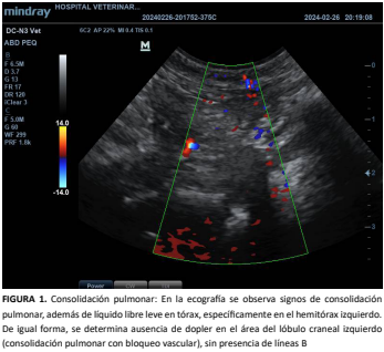
Torsión de lóbulo pulmonar craneal izquierdo en un perro diagnosticado mediante imágenes: Reporte de caso.
Resumen
La torsión pulmonar en perros, y de forma particular en razas braquicéfalas, se presenta como una patología compleja y potencialmente letal en caninos, la cual se caracteriza por una rotación anormal de un lóbulo pulmonar alrededor de su hilio. Esta condición compromete la vascularización y drenaje linfático del lóbulo afectado, induciendo isquemia, edema y, eventualmente, necrosis tisular. Los signos clínicos son a menudo inespecíficos y pueden confundirse con otras enfermedades respiratorias, dificultando su diagnóstico temprano. En este estudio se presenta el caso de un perro de raza Pug que fue atendido por un cuadro clínico compatible con enfermedad respiratoria, y en el cual mediante la realización de una tomografía computarizada (TC), se diagnosticó con torsión del lóbulo pulmonar craneal izquierdo. La TC se mostró como una herramienta diagnóstica esencial para visualizar las alteraciones anatómicas características de esta patología y permitiendo además descartar otros diagnósticos diferenciales. Ante la gravedad del cuadro clínico, se realizó una lobectomía pulmonar. Al presentar el paciente una evolución postoperatoria favorable, se resalta la importancia de un diagnóstico temprano y un manejo quirúrgico oportuno para mejorar el pronóstico. De igual forma se debe considerar que la torsión pulmonar representa un desafío diagnóstico y terapéutico, por lo cual la TC debe considerarse como el examen de elección para confirmar el diagnóstico y evaluar la extensión de la lesión, de igual forma el tratamiento quirúrgico, aunque complejo, ofrece excelentes resultados en estos pacientes.
Descargas
Citas
Rubin JA, Green J. Lung Lobe Torsion. En: Aronson LR, ed. Small Anim Surg Emergencies. 2nd Ed. John Wiley & Sons, Inc; 2022. p 451–458. doi: https://doi.org/pbr5 DOI: https://doi.org/10.1002/9781119658634.ch39
Lee T, Nam A, Lee DK, Lee HJ, Song KH. Lung lobe torsion in a dog with a tracheal stent for severe tracheal collapse. Korean J. Vet. Serv. [Internet]. 2023; 46(4):349–355. doi: https://doi.org/pbr6 DOI: https://doi.org/10.7853/kjvs.2023.46.4.349
Park KM, Grimes JA, Wallace ML, Sterman AA, Thieman-Mankin KM, Campbell BG, Flannery EE, Milovancev M, Mathews KG, Schmiedt CW. Lung lobe torsion in dogs: 52 cases (2005–2017). Vet. Surg. [Internet]. 2018;47(8):1002–1008. doi: https://doi.org/pbr7 DOI: https://doi.org/10.1111/vsu.13108
Lee SK, Cho KO, Alfajaro MM, Lee J, Yu D, Choi J. Use of computed tomography and minimum intensity projection in the detection of lobar pneumonia mimicking lung lobe torsion in a dog. Vet. Radiol. Ultrasound. [Internet]. 2019; 60(5):E48–53. doi: https://doi.org/pbr8 DOI: https://doi.org/10.1111/vru.12565
D’Anjou MA, Tidwell AS, Hecht S. Radiographic diagnosis of lung lobe torsion. Vet. Radiol. Ultrasound. [Internet]. 2005; 46(6):478–484. doi: https://doi.org/b5qg8p DOI: https://doi.org/10.1111/j.1740-8261.2005.00087.x
Hareardóttir H, Thierry F, Murison PJ. Anaesthesia management of a pug (in late-stage pregnancy) with lung lobe torsion. Vet. Rec. Case Reports. [Internet]. 2019; 7(2):e000765. doi: https://doi.org/pbr9 DOI: https://doi.org/10.1136/vetreccr-2018-000765
Belmudes A, Gory G, Cauvin E, Combes A, Gallois-Bride H, Couturier L, Rault DN. Lung lobe torsion in 15 dogs: Peripheral band sign on ultrasound. Vet. Radiol. Ultrasound. [Internet]. 2021; 62(1):116–125. doi: https://doi.org/pbsb DOI: https://doi.org/10.1111/vru.12918
Ciriano E, Marrington M, Grant J. Lung lobe torsion in association with a pulmonary papillary carcinoma in a dog. J. S. Afr. Vet. Assoc. [Internet]. 2022; 93(2):160–163. doi: https://doi.org/pbsc DOI: https://doi.org/10.36303/JSAVA.515
Gall N, Butts DR, Chanoit GP, Major AC. Computer tomography measurements of the airway and thoracic cavity do not provide support for bronchial conformation as a predisposing factor of left cranial lung lobe torsion in pugs. Vet. Radiol. Ultrasound. [Internet]. 2024; 65(3):255–263. doi: https://doi.org/pbsd DOI: https://doi.org/10.1111/vru.13345
Sumping JC, O’connell EM, Mortier J. Computed tomographic and clinical findings in a dog with suspected liver lobe torsion, secondary disseminated intravascular coagulation and multiorgan infarction. Vet. Rec. Case Reports. [Internet]. 2020; 8(4):e001166. doi: https://doi.org/pbsf DOI: https://doi.org/10.1136/vetreccr-2020-001166
Epstein SE, Balsa IM. Canine and Feline Exudative Pleural Diseases. Vet. Clin. North Am. Small Anim. Pract. [Internet]. 2020; 50(2):467–487. doi: https://doi.org/pbsg DOI: https://doi.org/10.1016/j.cvsm.2019.10.008
Howes CL, Sumner JP, Ahlstrand K, Hardie RJ, Anderson D, Woods S, Goh D, de la Puerta B, Brissot HN, Das S, Nolff M, Liehmann L, Chanoit G. Long-term clinical outcomes following surgery for spontaneous pneumothorax caused by pulmonary blebs and bullae in dogs – a multicentre (AVSTS Research Cooperative) retrospective study. J. Small. Anim. Pract. [Internet]. 2020; 61(7):436–441. doi: https://doi.org/pbsh DOI: https://doi.org/10.1111/jsap.13146
Elliott RC, Cassel N. Chronic lung lobe torsion in a pug. Vet. Rec. Case Reports. [Internet]. 2018; 6(3): e000662. doi: https://doi.org/pbsj DOI: https://doi.org/10.1136/vetreccr-2018-000662
Davies JA, Snead ECR, Pharr JW. Tussive syncope in a pug with lung-lobe torsion. Can. Vet. J. [Internet]. 2011; 52(6):656–660. PMID: 22131584. https://n9.cl/dvifex
Hansen NL, Hall SA, Lavelle R, Christie BA, Charles JA. Segmental lung lobe torsion in a 7-week-old Pug. J Vet Emerg Crit Care. [Internet]. 2006;16(3):215–218. doi: https://doi.org/dsdkh2 DOI: https://doi.org/10.1111/j.1476-4431.2005.00179.x
Latimer CR, Lux CN, Sutton JS, Culp WTN. Lung lobe torsion in seven Juvenile dogs. J. Am. Vet. Med. Assoc. [Internet]. 2017; 251(12):1450–1456. doi: https://doi.org/gcqgsv DOI: https://doi.org/10.2460/javma.251.12.1450
Felson B. Lung torsion: radiographic findings in nine cases. Radiology. [Internet]. 1987; 162(3):631–638. doi: https://doi.org/pbsk DOI: https://doi.org/10.1148/radiology.162.3.3809475
Tamburro R, Pietra M, Militerno G, Diana A, Spadari A, Valentini S. Left cranial lung torsion in a bernese mountain dog: A case report. Vet. Med. (Praha) [Internet]. 2011; 56(8):416–422. doi: https://doi.org/pbsm DOI: https://doi.org/10.17221/1553-VETMED
Lee KJ, Choi SJ, Kim YH, Jeong IS, Choi HJ, Lee YW. Radiography and computed tomography in four dogs with lung lobe torsion. J. Vet. Clin. [Internet]. 2013; 30(5):390–393. Disponible en: https://goo.su/ibxRxTc
Jeong S, Seo J, Lee J, Chang HS, Choi M, Yoon J. Imaging features of lung lobe torsion in two dogs with typical or atypical initial radiographic signs. J. Vet. Clin. [Internet]. 2018; 35(6):282–285. doi: https://doi.org/pbsn DOI: https://doi.org/10.17555/jvc.2018.12.35.6.282


